Day 1 :
- Case Reports on Psychology |Neurology| Ophthalmology |Dentistry |Cardiology |Pulmonology |Gastroenterology |Diabetes |Obstetrics and Gynaecology |Epidemiology |Surgical | Paediatrics| Public Health | Dermatology |Emergency Medicine and Critical Care |Forensic and Legal Medicine |Internal Medicine |Orthopaedics & Rheumatology |Pharmacology and Therapeutics |Women’s Health |Radiology |Anaesthesiology |Pathology- Anatomic & Clinical |Sexual Health |Cancer Science| Clinical Pathology |Geriatric Medicine |Veterinary |Vascular and Endovascular Surgery
Session Introduction
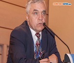
Sergey Suchkov
I M Sechenov First Moscow State Medical University, Russia
Title: A novel biomarker-based tool to monitor multiple sclerosis evolution at clinical and sub-clinical stages: Antibody-proteases as a new generation of translational tools to monitor, to predict and to prevent demyelination
Time : 09:00-09:30

Biography:
Sergey Suchkov is a Researcher-Immunologist, a Clinician graduated from Astrakhan State Medical University, Russia in 1980. He has been trained at the Institute for Medical Enzy-mology, The USSR Academy of Medical Sciences, National Center for Immunology (Rus-sia), NIH, Bethesda, USA and British Society for Immunology to cover 4 British university facilities. Since 2005, he has been working as Faculty Professor of I.M. Sechenov First Mos-cow State Medical University and of A.I. Evdokimov Moscow State Medical & Dental Uni-versity. He is the First Vice-President and Dean of the School of PPPM Politics and Man-agement of the University of World Politics and Law. He was a Scientifi c Secretary-in-Chief of the Editorial Board of the International Journal “Biomedical Science” (Russian Academy of Sciences and Royal Society of Chemistry, UK) and The International Publishing Bureau at the Presidium of the Russian Academy of Sciences. He was a Director of the Russian-American Program in Immunology of the Eye Diseases. He is a Member of EPMA, NY Academy of Sciences and an Editorial Board Member for Open Journal of Immunology and others.
Abstract:
Subclinical multiple sclerosis (S-MS) can be usually defined as the discovery of characteristic lesions at magnetic resonance (MR) or at autopsy, in the absence of clinical evidence con-sistent with MS. The methodological bricks of subclinical diagnostic and predictive protocols should include basic algorithms to differ essentially from those employed in canonical clinical practice, i.e., (i) to confirm a diagnosis of subclinical stage of the disease course and (ii) to select a mode for preventive treatment to quench the autoimmune inflammation. In this sense, among the best-validated proteome-related translational biomarkers, antibody-proteases were proven to be the best known ones. Abs against myelin basic protein/MBP endowing with proteolytic activity (Ab-proteases with functionality) is of great value to monitor demyelination to illustrate the evolution of MS. The activity of the MBP-targeted Ab-proteases discovered in MS patients markedly differs between: (i) MS patients and healthy controls; (ii) different clinical MS courses; (iii) EDSS scales of demyelination to correlate with the disability of MS patients to predict the transformation prior to changes of the clinical course. The activity of Ab-proteases was first registered at the subclinical stages 1-2 years prior to the clinical illness. About 24% of the direct MS-related relatives (probands) were se-ropositive for low-active Ab-proteases from which 38% of the seropositive relatives estab-lished were being monitored for 2 years whilst demonstrating a stable growth of the Ab-associated proteolytic activity. Three patients were initially evaluated because of accidental MRI findings suggestive of MS that fulfilled the Barkhof criteria. At the moment of MR ex-amination, patients were asymptomatic. The objective examinations as well as the clinical history were negative. After having those patients tested for Ab-proteases, all three have demonstrated elevated levels of the specific activity to target MBP. We have been monitoring along with the patients mentioned all direct members (13 healthy persons) of their families for two years and found that three relatives tested had elevated levels of the specific activity which was having a trend to grow whilst correlating with clinical symptoms of MS including the chronic fatigue, muscle weakness, dizziness, etc. All family members were studied with MRI, evoked potentials, and human leukocyte antigen (HLA) typing. The activity of Ab-proteases and its dynamics tested would confirm a high subclinical and predictive (transla-tional) value of the tools as applicable for personalized monitoring protocols. Further studies on targeted Ab-mediated proteolysis may provide a translational tool for predicting demye-lination and thus the disability of the MS patients in a variety of clinical and subclinical cases.
Nick Kostovic
Kostovic Acupuncture by bio Electron’s Laser, USA
Title: Cancer cured by bio electron's laser acupuncture
Time : 09:30-10:00

Biography:
Nick Kostovic was graduated from the Split Gymnasium in 1969 with an Associate of Art’s degree in Humanities and Science. He has combined Quantum Mechanics Physics and Electromagnetic Science and created K-BTE Medical Laser Device. He has canceled magnetic from industrial electromagnetic electricity and converted this into his discovery bioelectricity which has the ability to release bio electron’s photons which become most efficient tool in healing process of any of human physical organs.
Abstract:
New Advanced Technology in Medicine by Bio Electron’s Photons Special circuit: I, Nick Kostovic, for the first time in recorded history have eliminated magnetic from regular electromagnetic electricity. I also created the next six steps described below. I did this by developing a proprietary way of reversed current RC to create what is bio electricity. The device I created is called the Kostovic BioTechnological Energizer, K-BTE Medical Laser Device First, my center has successfully developed special current circuit and canceled magnetic from electromagnetic. Second, this device extracts bio electron photons from H2O electric fluid by wire and wirelessly. Third, while using the K-BTE device therapist has absolute control of speed/frequency of these released and enriched bio electron photons. Fourth, bio electrons photons are converted into the strength of Micro or Nano amperes allowing the bio electricity to softly penetrate into the brain or any other physical organ with zero harm to the healthy cells. Fifth, in the process of extracting bio electron photons from the electric fluid it can include transference of hundreds of different natural acids as well as amino acids. Each biological agent BA is capable to transfer 3 to 6 different natural and amino acids, by enriched bio electron photons Sixth, these enriched bio electrons photons are wirelessly transferred through and olive oil coating on the skin enabling the bio electricity to softly penetrate/bio electron photons always penetrate softly on the skin surface/deeply and efficiently targeting the specific ailing human tissue. This process is always skillfully directed into the body with the very gentle frequencies of Micro and Nano amperes allowing zero risk of negative consequences.
Milen Velinov
NYS Institute for Basic Research in Developmental Disabilities, USA
Title: Genomic medicine empowers personalized patient care-an example of effective albuterol treatment for a patient with homozygous DOK7 mutation
Time : 10:00-10:30

Biography:
Milen Velinov is an Associate Professor at Albert Einstein College of Medicine and Director of the Genetic Service at Bronx-Lebanon Hospital. He is also the Program Director of Comprehensive Genetic Services and Specialty Clinical Laboratories at The New York State Institute for Basic Research in Developmental Disabilities. His research interests include Neuronal Ceroid Lipofuscinoses as well as rare/unique clinical manifestations and diagnoses in clinical genetics.
Abstract:
A 15 years old female patient was referred for genetic evaluation because of dysarthria and muscle hypotonic. Her early motor development was delayed. She did not start walking until 2.5 years. Reportedly her early speech development was not delayed. However at her first visit with us she was noted to have dysarthria. She was also reported to have academic difficulties, possibly associated with her dysarthria. This patient had prominent muscle weakness. She had great difficulty getting upright from squatting position. She could not run and sometimes had impaired balance. The patient’s parents were first cousins. Screening laboratory tests and molecular testing for Friedreich ataxia were normal. Whole exome sequencing was done because no specific etiology was apparent. The patient tested positive for homozygous pathogenic sequence change in gene DOK7 (c.1124_1127dupTGCC). Homozygous mutations in this gene were previously associated with congenital myastenic syndrome type 10 (CMS10). Previously reported Albuterol treatment in 15 patients with DOK7 mutations led to improvement in these patients’ muscle strength. Treatment of our patient was initiated with 4 mg P.O. Albuterol daily. She initially developed borderline increase in her blood pressure and the Albuterol dose was decreased to 2mg alternated with 4 mg daily. At this regimen no adverse effects were observed. Monthly fatigue tests examining the muscle strength were conducted and a significant improvement of muscle strength of hands, abdominal muscles and legs was recorded. In addition the patient’s parents reported improvement in her speech. The patient continues to receive Albuterol treatment. Personalized medicine is a relatively new developing medical approach that already has wide application in cancer care and clinical genetics. This new field was greatly empowered by the development of technologies for massive nucleic acid sequencing. Identifying specific
Ehab M. Esheiba
Thumbay Hospital, UAE
Title: Chronic thrombo-embolic pulmonary hypertension (CTEPH)
Time : 10:30-11:00

Biography:
Ehab M Esheiba has completed his Postgraduate Diploma in Internal Medicine and Master Degree in Cardiology in 2004. Later he completed his MRCPUK and he is a Fellow of the Royal College of Physicians of Edinburgh. He is a Member of the European Society of Cardiology and European Association of Cardiovascular Imaging. He has several publications and presentations at national and international levels. He is currently involved in several clinical research projects as a Principal Investigator and also as a Supervisor and Co-Supervisor. He is an Editorial Board Member of a reputed cardiology journal. Currently, he is working as Head of the Cardiology Department of Thumbay Hospital, Gulf Medical University, Ajman, UAE.
Abstract:
Chronic thrombo-embolic pulmonary hypertension (CTEPH) is defined as an elevation of the mean pulmonary arterial pressure above 25 mmHg that persists six months after an episode of pulmonary embolism (PE). It occurs an about 2-4% in survivors from PE. The clinical picture, natural history and prognosis may vary from one patient to another. Venous thromboembolism (VTE), including both deep vein thrombosis (DVT) and PE is the third most common cardiovascular illness after acute coronary syndrome and stroke. A 29 year old male patient presented with gradually progressive shortness of breath and fatigue for several months. He had one episode of loss of consciousness. Clinical examination revealed picture of pulmonary hypertension. Further laboratory and imaging confirmed the presence of recent pulmonary embolism and markedly elevated pulmonary arterial pressure, consistent with the diagnosis of CTEPH. In this article, we discussed this clinical case, the diagnostic pathway and the management plan that was followed with the patient till his last follow-up, focusing on guidance on management in the same context. We believe that addressing VTE as a possible regional public health problem should take a multi-dimensional approach targeting the epidemiology of the disease with implementation of cost-effective preventive and therapeutic programs.
Maria Voyatzi
Dermatologist – Venereologist, Greece
Title: Efficacy of lasers on pigmentary lesions,telangiectasias , hemangiomas and rejuvenation

Biography:
Maria Voyatzi has completed her PhD from the Aristotle University of Thessaloniki, Greece and postdoctoral studies from the Hospital of Dermatological and Venereological Diseases of Thessaloniki, State Clinic. She has published more than 15 papers in reputed journals and participated in many Hellenic, European and world congresses.
Abstract:
Our purpose is to study the efficacy of the Alexandrite laser (755 nm) and the fractional laser when treating pigmentary lesions, the efficacy of the NdYAG laser (1064 nm) when treating telangiectasia and hemangiomas and the efficacy of the NdYAG laser and fractional laser on rejuvenation. We used the data from our private office from the past five years (2011-2015). We treated 1000 patients with freckles with Alexandrite laser, 200 patients with melasma with fractional laser, 100 patients with postinflammatory hyperpigmentation with Alexandrite laser and 60 patients with postinflammatory hyperpigmentation with fractional laser. We also studied the side effects of the therapies. Alexandrite laser had excellent results on freckles, while the combination of chemical peels and fractional laser was satisfactory for the treatment of melasma. Post inflammatory hyperpigmentation had an intermediate response. The recurrence rates were higher in melasma and the side effects were generally minimal. We also treated 500 patients with telengiectasias and 300 patients with hemangiomas with NdYAG laser. As far as telangiectasias were concerned, the results were much better when the face was treated. In contrast, the recurrence rates were much higher when the legs were treated. The results were impressive in almost all cases of cherry hemangiomas. The combination of NdYAG lasers and fractional lasers on a monthly basis had satisfactory results in rejuvenation.

Biography:
Ali Moghimi is graduated from the Iran University of Medical Sciences, Tehran, with his medical degree in 2002. He worked as both a General Practitioner and Research Assistant for few years in Iran before moving to Australia. He began his specialty training in Anatomical Pathology in 2009 and obtained his Fellowship of the Royal College of Pathologists of Australasia early in 2014. He is currently a Member of the International Academy of Pathology and has over 10 publications to his name. He joined Dorevitch Pathology in 2014 and his special interests are obstetrics and gynaecological, perinatal and renal pathology.
Abstract:
Bartholin gland cyst results from dilatation of the Bartholin duct due to obstruction. Pigmentation in an external genital cyst, such as median raphe cyst, has been previously reported. However, presence of melanocytes or melanin pigment in the cyst wall of Bartholin duct cysts is an extremely rare finding, with only one case reported in the literature. Herein, I report the second case of pigmented Bartholin duct cyst with a brief review on pigmented cystic lesions in anogenital area. An 18-year-old lady presented with small pigmented papule in vulva, clinically resembling haematoma. The biopsy identified squamous mucosa with underlying stroma showing cystic spaces, lined by single or double layers of cuboidal to flat epithelium. Some epithelial cells contained brown pigment granules in their cytoplasm and there were also pigments within the surrounding stroma as well as the lumina of cystic spaces. The melanocytes within the epithelium of the cyst were stained by Melan-A, SOX10, MIT1 and S100 immunoperoxidase stains. A diagnosis of pigmented Bartholin duct cyst was rendered. This is the second documented case of pigmented Bartholin duct cyst in the literature. The first was a 41-year-old Japanese female who presented with an asymptomatic nodule in the external genitals, showing features similar to this case. The mechanism of pigmentation has not been elucidated and there are no malignant features associated with this finding. However, it is important for reporting pathologist to be aware of this variant in order to avoid misinterpretation of the findings as an atypical melanocytic lesion.
Hind Manaa Alkatan
King Saud University, Saudi Arabia
Title: Periorbital nodular fasciitis in an adult male: The first reported case in the Middle East with review of the literature

Biography:
Hind Manaa Alkatan has completed her Saudi Ophthalmology Board from King Saud University (KSU), Riyadh, KSA and her postdoctoral studies from Departments of Ophthalmology/Pathology, University of Manitoba and University of British Columbia, Canada. She is an Assistant Professor, Consultant (Departments of Ophthalmology and Pathology), Chief of Ophthalmic Pathology, and Director of the Post-Graduate Residency and Fellowship Training Programs in Ophthalmology, King Saud University Medical City (KSUMC), Riyadh, KSA. She is a Member in international organizations: Eastern Ophthalmic Pathology, Canadian Ophthalmology Society, International Society of Ocular Oncology, Ocular Oncology Group in addition to the local Saudi Ophthalmology Society.
Abstract:
Objectives: Nodular fasciitis (NF) is a benign, fibroblastic proliferation, which can raise suspicion for malignancy and is relatively rare in the ocular region. We report the first case of periorbital NF in the Middle East with review of the literature.
Methods: Our case of NF is described clinically and histopathologically in details. An extensive review of all reported cases of ocular/ periocular NF in the English-written literature is made with summary of 39 cases including ours taking into consideration the location of the tumor, age and gender.
Results: Our 35-year-old male presented with a recurrent tumor in the periocular region. Diagnosis of NF was made by careful histopathological examination of the excisional biopsy and by immunohistochemical staining. We have summarized the demographics and tumor location of 38 cases previously reported and added our case information.
Conclusions: Demographically, NF occurs within a wide range of age (8 months to 81 years) with a mean age of about 28 years. Half of the cases are in the age group of 30 years or younger (median=30 years). Slight female predominance is observed (Female : Male ratio is =7:6). The right side is more commonly involved (in 21/37). Periocular/orbital involvement is more commonly found (67%).
Valsamaki P
General Hospital of Athens Alexandra, Greece
Title: Uterine leiomyomas in the differential diagnosis upon 99mTc-EDDA/HYNIC-TOC interpretation

Biography:
Valsamaki P has completed her PhD from Aristotle University School of Medicine of Thessaloniki, Greece and postgraduate studies from Aristotle University of Thessaloniki, National and Kapodistrian University School of Medicine of Athens, Greece, and Nuclear Medicine Department, University of Bologna, Italy. She works as consultant in the Nuclear Medicine Department of the UGHospital Alexandra, Athens, Greece. She has published more than 25 papers in reputed journals, participated in more than 10 research protocols, received highlights distinction in 10 presentations/articles, and has been serving as an Editorial Board Member (HJNM) and as a reviewer (ANM) of repute.
Abstract:
The diagnostic algorithm of neuroendocrine tumours encompasses somatostatin receptor scintigraphy as a useful non-invasive method which shows high sensitivity and specificity. This technique also indicates the option of targeted therapeutic “cold” or “hot” (radiolabeled) somatostatin analogue application. We present three cases of uterine leiomyomas with or without uterus dislocation, identified by 99mTc-EDDA/HYNIC-TOC (Tektrotyd) in women of different ages. Images obtained 75 min up to 240 min after injection of 740 MBq of 99mTc-labeled EDDA/HYNIC-TOC show areas of abnormally increased uptake in pelvis, with or without bladder compression. Our cases indicate that somatostatin receptors may be present in the uterus and in leiomyomas, irrespective of age. The findings were confirmed by other imaging modalities, such as US, CT and/or MRI. Uterine leiomyoma(s) somatostatin receptor expression proposes the potential utilization of somatostatin analogues in the non-invasive treatment armamentarium of these disease entities. Limited literature reports, compatible with our observations, highlight the significance of this finding in the differential diagnosis of respective positive studies.

Biography:
Nicolas Martinelli is a student at the faculty of medicine and surgery of the University of Pavia. At present, he has published one paper in the Journal of pediatric
care.
Abstract:
We describe the case of an Italian, Caucasian nine-year-old girl, suffering from a unilateral tonsillar swelling. Histopathological and immune-histochemical exams show a lymphoepithelial reactive hyperplasia overlap with a chronicle inflammation. Microbiologic swaps reveal a throat infection caused by Streptococcus pyogenes, and a probable viral infection, caused by Ebstein Barr Virus (EBV). We underline the importance to recognise and consider this benign lesion due to its similarities with a malignant neoplastic form, especially during differential diagnosis of cervical swellings in young women. This is one of the rare cases described in scientific literature in the last years, especially among caucasian race.

Biography:
Amer Hashim Al Ani currently works as a general and GIT surgeon in the department of general surgery at Sheikh Khalifa Hospital, United Arab Emirates
Abstract:
Introduction: The inguinal canal is a 3.75 cm passage in the anterior abdominal wall. It seems too small to miss or to find a foreign body in it. Foreign bodies can reach the inguinal canal in different ways; by ingestion, from the lumen, left by a surgeon, or through the abdominal wall. They may migrate from the peritoneal cavity e.g., gall stones spilled free during laparoscopic surgery. Foreign bodies may include fish, chicken bones, tooth picks, needles, safety pins, piece of wood, glass, sponge and gauze .The gastrointestinal tract is the most common route by which foreign bodies can reach the inguinal canal.
Case study: We present her two patients, both were males and their age was 49 and 70 years. The first (With a history of CVA on Aspirin) was presented to ER with signs and symptoms of intestinal obstruction due to a suspicious strangulated inguinal hernia. The
second (A case of chronic renal failure on hemodialysis) was presented to surgery clinic with history of chronic scrotal sinus on the same side of recurrent inguinal hernia. Both underwent exploration of the inguinal canal.
Result: Exploration of 1st inguinal canal revealed a balloon of a Foley's catheter inside a diverticulum of urinary bladder, which was a part of sliding inguinal hernia (the cause of intestinal obstruction was diagnosed as superior mesenteric artery occlusion during same
session laparotomy). Exploration of 2nd inguinal canal revealed missed surgical gauze left during the previous hernia repair.
Conclusion: The presence of foreign bodies in the inguinal canal is rare. Still they can be found during exploring the inguinal canal for hernia repair.
V I Petukhov
Vladimir State University, Russia
Title: Electrogenic metals in epidermis and the synchronous operation of membrane ATPases

Biography:
V I Petukhov currently works in the field of psychology in the Vladimir State University, Russia.
Abstract:
The homeostasis of electrogenic metals (K, Na, and Ca) in epidermal cells can be studied using a derivative of epidermis (hair). This approach was used in the present study to confirm the close relationship between electrogenic metals (EM) and cell bioenergetics and find evidence for possible attributability of EM homeostasis to the phenomena of self-organized criticality (SC). The above assumption is based on the following well-known facts: Generation and maintenance of electrochemical potential of the cell at the expense of permanent and multi-directional EM traffic across the plasma membrane; Participation of sodium ions (Na+) in cellular energy exchanges as a convertible ‘energy currency’, complementary to adenosine triphosphate (ATP); Failure of the hypothesis of normal distribution of quantitative spectrometry data of metals contained in epidermis derivatives (hair). Using mathematical statistics, we have analyzed the results of atomic emission spectrometry of hair samples for Na, K, and Ca content, which were obtained at the Center for Biotic Medicine (Moscow) from 10297 healthy subjects (5160 males and 5137 females) aged 2 to 85. Our previous studies have found that the results of quantitative spectrometry of EM in epidermal cells (hair) from healthy individuals and the liquidators of the chernobyl accident (chronic oxidative/nitrosative stress) are conjugated. The nature of this conjugation, like many intimate mechanisms of the metal-ligand homeostasis in the epidermis, remains unsolved. The results led to the following conclusions: Normal functioning of membrane Na+/K+-ATPases suggests synchronous (critical) nature of transmembrane traffic for EM. The synchronous operation of membrane ATPases is confirmed by the positive K-Na relation (Pearson) and signs of self-organized criticality (SC) according to the data of mathematical analysis and the obtained results point to the probable attributability of EM homeostasis in epidermis to SC-phenomena.
Paulo Eduardo Ocke Reis
Fluminense Federal University, Brazil
Title: Endovascular treatment of focal infrarenal aortic stenosis with absence of the celiac trunk - case report

Biography:
Reis PEO is specialist in Vascular Surgery and Endovascular Surgery . Adjunct Professor of Vascular Surgery and Professor, Graduate Course in Cardiovascular Sciences. He is coordinator of the Vascular Surgery Service, University Hospital Antonio Pedro, Federal University Fluminense. Brazil / Rio de Janeiro / Niterói .He is also Editor-in-Chief-Journal of Vascular & Endovascular Surgery . EJVES Short Reports Editorial Board - Associate Editor
Abstract:
This case report describes a case of abdominal aortic stenotic disease treated with covered balloon- expandable stent with absence of the celiac trunk artery. Technique: Patients are selected for this strategy if they have a lesion without bifurcation involvement located at the mid segment of the infrarenal aorta or stenosis with unfavorable aortic anatomy, or severely diseased and calcified distal aorta. Conclusion: Our result in this case supports the feasibility, safety, and efficacy of stenting for stenosis of the distal infrarenal aorta.
Keywords: aortic stenosis, endovascular procedures, angioplasty, endovascular surgery, celiac trunk variation
Brian Zeman
Ryde and Royal North Shore Hospitals, Australia
Title: Case report- Psychosis due to iatrogenic causes, cerebral palsy or spontaneous

Biography:
Brian Zeman is a medical specialist in Rehabilitation Medicine practising in Sydney. He completed his medical degree at UNSW and then specialist training with the ACRM becoming one of the first four graduates. The ACRM later became a Faculty in the RACP. He has presented papers overseas on various subjects and is involved in medical student teaching as Lecturer. As well he is Clinical Supervisor for specialist training in rehabiliation medicine. He has interests in neurological rehabilitation, burns rehabilitation and medicolegal reports.
Abstract:
Ms M is a 42 year old woman with cerebral palsy affecting her mobility and speech. She was independent in the community and working. She also had unrelated chronic asthma and after an exacerbation was prescribed prednisone. She then developed psychosis gradually over four weeks. The prednisone was stopped and she was prescribed an antipsychotic. She then developed extrapyramidal reaction causing torticollis and limb spasm. The antipyschotic was ceased and she was prescribed Benztropine. She did not improve and remained hospitalised. She had Botulinum toxin injections to the neck and later leg as well as extensive physiotherapy with gradual improvement. There was some improvement but not to previous mobility levels. About two monhts later, she again gradually developed psychosis with auditory and visual hallucinations. She had extensive investigations but there were no obvious causes. Management of the psychosis with medications has been difficult and she required admission to psychiatric unit to have these monitored. Was the psychosis due to triggered by prednisone, related to her CP and immature brain or was it spontaneous and unrelated?
Edgar R. Abascal-Garcia
Intensive Care Unit Hospital, Mexico
Title: Early use of continuous renal support therapy in HELLP syndrome

Biography:
Dr. Abascal-Garcia completed the Emergency Medicine program and Critical Care specialization at the National Autonomous University in Mexico. He is part of the Intensive Care Staff at Hospital Espanol, a renowned institution in Mexico. He has published several papers in reputed national journals focusing in Intensive Care.
Abstract:
Case: A 29 years old obese and diabetic pregnant woman (29 weeks of gestation) presented to the emergency room with 200/120 of blood pressure nonresponsive to oral medication nor sulfate magnesium infusion. Laboratories and other vasospasm manifestations confirmed an overt HELLP syndrome. She underwent an emergency C-section and was admitted into the Intensive Care unit (ICU) afterwards. She was started on intravenous Labetalol and mechanical ventilation due to respiratory failure. She evolved with oliguria (KDIGO Stage 1) despite of a fluid accumulation of 6% in a 6-hour period. Despite hemodynamic stability was achieved and renal biomarkers being unaltered, continuous renal replacement therapy (CRRT) was initiated in a full convection modality. Clinical and biochemical normalization occurred after 48 hours and mechanical ventilation was withdrawn. She was disconnected from renal support 24 hours after extubation, and discharged from the ICU 72 hours after the admission. Urinary flow remained adequate during the whole ICU stay. She and her baby were discharged healthy to home.
Discussion: Proinflammatory Cytokines damaging the endothelium is part of the still not fully understood HELLP syndrome pathogenesis. These cytokines (IL-2, TNF) have a molecular weight compatible with convection clearance. CRRT has been used without severe kidney injury in Acute Respiratory Distress Syndrome, pancreatitis and other syndromes with good outcomes. The early use of CRRT might have accelerated patient´s recovery.
Conclusion: CRRT exhibited benefit in a case of HELLP syndrome with KDIGO stage I AKI. This pathology should be explored as a non-renal indication for CRRT.
Thamer ALOhali
King Fahad Medical City, KSA
Title: Improvement in diabetic patient immunization at the family medicine department, KFMC

Biography:
Thamer AlOhali is a Family Medicine Consultant with subspecialty in public health and healthcare quality improvment. He is the Chairman of Family Medicine Department as well as the Chairman of Family Medicine Quality Improvement Committee at KFMC. He has completed his Residency Training program at Prince Sultan Military Medical City in Riyadh on 2011. He obtained his Clinical Health Care Quality and Patient Safety Fellowship in University College London Hospitals on 2014 and, on January 2016 he completed his Clinical Fellowship in Primary Care and Public Health at Imperial College London.
Abstract:
Diabetic patients are immune-compromised patients and they are at high risk to be infected. Thus, the American Diabetes Association (ADA) recommends Influenza, Pneumococcal, and Hepatitis B vaccines for all diabetic patients to reduce the risk of morbidity and mortality. Diabetic Patients in the Family Medicine Department at KFMC were not properly provided with the recommended immunizations according to the ADA guideline which affects patient satisfaction and well-being. The project team has been organized to assess the problem and analyze its causes. During 2015, only about 10% of diabetic patients have been offered the recommended vaccines by their healthcare workers. There are many factors behind this. The most important factors are: Patient overload, lack of reminder, insufficient education about the importance of immunization, patient compliance and unavailability of a clear pathway. In order to ensure that all diabetic patients visiting the department have the chance to be educated and offered the vaccines, a design of pathway for improvement in Diabetic Patient Immunization has been created. The goal was to increase the proportion of diabetic patients who receive recommended immunizations based on the ADA guidelines from about 10% to 60% by December 2016. The compliance rate for improvement in diabetic patient immunization in overall reached about 60%. Starting by August 2016, using data registry, the average ranging for August 2016 was 36%, while 68% for the month of September 2016 and 60% for the month of October 2016 which reached our target compliance rate of 60% for the year 2016.
Youssef Abo Elwan
Hera General Hospital, Saudi Arabia
Title: Pregnancy with anemia and severe thrombocytopenia secondary to vitamin b12 deficiency: a rare presentation

Biography:
Youssef Abo Elwan works as a Professor of Obstetrics & Gynecology, Faculty of Medicine, Zagazig University, Egypt. He is the head of High Risk Pregnancy Unite. Faculty of Medicine; Zagazig University, Egypt. He is the member of High council Supreme for Professor Promotion in Egyptian Universities. He is also the Reviewer in many national & international journals.
Abstract:
A 24 years old Saudi Patient G3 P2 +0,35 weeks pregnancy, was admitted as an emergency case complaining of dizziness and fatigability; she also suffered of nausea all over her pregnancy. She had history of delivery of anencephaly , she was a known case of hypothyroidism. Upon admission she was pale but not jaundiced. Her Hb was 4.9 gm/dl, MCV 82.30 fl (80-101), MCH 28.80 pg (27-33), Retics% 0.18 (0.2-2), her platelet count dropped from 40,000 to 27,000 /mm3 after 3 days from admission LDH 5175. Her B.P 120/80, no proteinurea, her ALT and AST was within normal. She received 3 units of packed RBCs on admission. Based on thrombocytopenia , high ESR and splenomegaly, she was through to have connective tissue disease and she received pulse methylprednisolone for 3 days, however with no response. Later on we repeated blood film and serum B12 level which showed hypersegmented neutrophil and serum B12 was very (31.9 pmol/L; N= 148-616). It was clear at that time, the cause of severe thrombocytopenia was vit B12 deficiency. So cyanocobalamine injection started which showed within few days marked improvement in platelet count up to 260.000/ mm3, Hb 11.7 g/dl, retics % 10.9, after less than 10 days from starting vit B12 injections. Patient continued her pregnancy and delivered spontaneously 2270 gm with good Apgar score. We concluded that vit B12 deficiency should be considered as a cause of severe thrombocytopenia although it is rare.
Jignesh Kothari
U N Mehta Institute of Cardiology and Research Center, India
Title: Coronary artery disease with coarctation of aorta in elderly patient

Biography:
Jignesh Kothari currently works in U. N. Mehta Institute of Cardiology and Research Center (affiliated to B J medical college, Ahmedabad), India
Abstract:
Coarctation of the aorta is typically a disease of childhood and early adulthood, and there is a reduced life expectancy in patients who have not undergone correction. Survival to older age is rare, due to severe cardiovascular complications. Because there are only a few cases of elderly patients with uncorrected aortic coarctation, management strategies in these patients are challenging. We describe the case, first diagnosed with Coronary Artery Disease, then developed aphasia, then diagnosed coarctation of aorta at 56 year of age. Coronary artery bypass grafting was performed, and the coarctation was managed with ventral aorta repair. At the six months follow-up visit, the patient was asymptomatic and maintained a normal blood pressure with minimal gradients between upper and lower extremities. His aphasia was cured after two month of surgery. To our best knowledge there is hardly such case report available in literature.

Biography:
Vasiliki E. Kalodimou MSc, Ph.D. is the director of the Flow Cytometry-Research and Regenerative Medicine Department at IASO Hospital in Athens, Greece. She has studied and worked with progenitor cells from placenta, umbilical cord, and adipose tissue along with their applications in regenerative medicine and flow cytometry, with publications in the field, including research fellowships. She has published 2 books about flow cytometry, the Greek edition was published in 2010 and in 2013 the book was published from AABB Press USA (Bestseller). In 2015 she was published her 3rd book, a handbook to mesenchymal stem cells in regenerative medicine by Specg.co
She is a Flow Cytometry/Stem Cell Specialist, Leader at AABB CT Subsection: CT Product Manufacturing and Testing-USA, Scientific Consultant in Regenerative Plastic Surgery Institute / Pure Aesthetics, Brazil, AABB Assessor-USA, editor in 12 scientific journals, editor-in-chief in 4 scientific journals, and her biography was included in Who Is Who in the world 2014, 2015 & 2016 edition.
Abstract:
The rejuvenation of their genitalia and the vagina will help to dramatically improve the quality of their lives.
Our aim is to prove that the infusion of mesenchymal stem cells contained in adipose tissue has an important contribution and long-term results:
1. In appearance and function of the skin and mucosa of the external female genitals and the vagina.
2.To reduce pain in women with dyspareunia
3. At the entrance of the vagina showing thinning of the epithelium and adhesions of granulation tissue.
4. To reduce urinary incontinence (from stress incontinence)
5. The improvement or treatment of lichen sclerosus of the vulva to restore the quality of life and sexual function in the affected women.
Osman A Hamour
King Faisal Specialist Hospital and Research Center, Saudi Arabia
Title: "Multiple Rare Complications Following Total Thyroidectomy and Bilateral Neck Dissection: Case Report and Literature Review"

Biography:
Osman A Hamour is currently working in the Department of General Surgery, King Faisal Specialist Hospital and Research Center, Saudi Arabia
Abstract:
Abstract Background: Thyroidectomy is a common and safe surgical procedure and is typically associated with low
morbidity and mortality. Delayed tracheal rupture after thyroidectomy has only infrequently been reported. Here we are
reporting a case in which a number of rare complications of total thyroidectomy and neck dissection happened over a
very short period of time with a review of existing literature.
Case presentation: A 48-year-old man, diagnosed as metastatic papillary thyroid cancer. Underwent total
thyroidectomy and bilateral modified neck dissection complicated by severe bronchospasm, bilateral pneumothorax,
pneumomediastinum, pneumopericardium managed by underwater seal drainage of his chest cavities. On the
10th postoperative day he developed spontaneous tracheal rupture which compromised his airway. He was coded,
eventually intubated resuscitated. In the operating room he had his neck explored, neck haematoma evacuated,
tracheal tear debrided and a tracheostomy tube was inserted.
Conclusion: Thyroidectomy remain a commonly performed safe surgery typically associated with low mortality
and morbidity. However other rare and serious complications as observed in this case can sometimes take plac and can be life threatening. Bilateral pneumothorax, pneumomediastinum and pneumopericardium are examples of such serious complications. Multiple contributing factors include heavy smoking, alleged barotrauma, and extensive electrocautery dissection. In our case the late spontaneous rupture of the trachea most likely been predisposed to by the excessive explosive smokers cough.
Luis Antonio R. Balajadia
Southern Philippines Medical Center Davao City, Philippines
Title: Primary adrenal hemangiopericytoma: The first reported case

Biography:
Luis Antonio R. Balajadia has completed his Doctor of Medicine at the age of 26 years from Davaao Medical School Foundation and 3 year General Surgery Residency Training at Davao Doctors Hospital. He is currently the Chief Resident of the Section of Urology in the Southern Philippines Medical Center.
Abstract:
Hemangiopericytomas are rare tumors originating from pericytes in the wall of capillaries. It is a type of soft tissue sarcoma which commonly involves the lower extremities, pelvic retroperitoneum, and head and neck. The age of initial diagnosis of HPC is 40.3 years-old (range 16-86) usually presenting as a painless mass. A 32-year old female presented with a 5 year history of slowly-growing right flank mass. She denied any history of hematuria, dizziness, headaches or hypertensive episodes. Physical examination findings showed a bulging right flank and an approximately 15 x 15 cm, palpable, nontender mass on bimanual examination. CT-scan with IV contrast was requested and revealed a large suprarenal mass, right. She underwent adrenalectomy with en-bloc nephrectomy, right with uneventful post-operative course. Histopathology of the specimen was read as hemangiopericytoma. Further testing by Flourescence In-Situ Hybridization confirmed the diagnosis. Metastatic work-up was done and was negative. Hence, this is the first reported case of primary adrenal hemangiopericytoma. Surgical removal is the mainstay of treatment of hemangiopericytomas. Radiotherapy and chemotherapy have no role in the management of the disease. The lesson learned from this case is that behind every mask there is a face, and behind that, a story. What seemed to be a simple and ordinary as an adrenal mass turned out to be as special and unique as a hemangiopericytoma.

Biography:
Mehmet Hüsamettin AKKÜÇÜK currently works in the Department of Emergency, Alanya Training and Medical Research Center, BaÅŸkent University Faculty of Medicine, Antalya, Turkey
Abstract:
Objective: Describing a case of a patient with presumed massive pulmonary embolism with
cardiac arrest and successfull resuscitation with fibrinolysis during CPR using rTPA.
Case summary: A 74-year-old man presented to the emergency departme for head trauma
due to syncope. On arrival, initial examination was suspect for myocardial infarction, and
cardiogenic shock but there were no signs of pulmonary overload. Cardiac ECHO showed a
dilated right heart and additional analysis turned the differential diagnosis to pulmonary
embolism.. Thrombolytic therapy was considered. His condition rapidly deteriorated and
progressed to cardiopulmonary arrest.During advanced cardiac life support, empirical
alteplase 50 mg was administered intravenously over 15 minutes with return of spontaneous
circulation (ROSC) The diagnosis of massive PE using computed tomography angiography
was confirmed after fibrinolytic therapy. During hospitalisation gastrointestinal bleeding
developed requiring transfusion, but no further complications occurred. Patient was
discharged after 15 days without any neurological sequelae.
Discussion: Diagnosing pulmonary embolism may be difficult. Massive pulmonary embolism
has a high mortality and early treatment is essential. Return of spontaneous circulation
(ROSC) is very rare in patients with cardiac arrest following massive PE. Clinical guidelines
recommend fibrinolytic therapy for patients with PE and cardiac arrest. It has classically been
contraindicated because of a concern for fatal bleeding. Current literature based on
retrospective studies and case reports however, seems to favor fibrinolytic treatment in PEassociated
cardiac arrest due to an increased chance of ROSC and improved survival.
CONCLUSION: Fibrinolysis may be beneficial in achieving ROSC, reducing mortality, and
preserving neurological function in PE-associated cardiac arrest patients, even in cases of
prolonged CPR and delayed fibrinolytic administration without increased risk
Jaewon Phee
Brighton and Sussex Medical School, UK
Title: An unusual presentation of Roth spots in Cogan’s Syndrome

Biography:
Jaewon has completed her B.Sc. (Hons) Medical Sciences from the University of Edinburgh in 2013 with an award for academic excellence. She is currently a 4th year medical student in Brighton and Sussex Medical School (BSMS) with research interest in ophthalmology. Her research output to date includes four national poster presentations. Jaewon is currently the committee member of the BSMS Ophthalmology Society and she is a member of Brighton Institute for Healthcare Improvement (IHI) Open School Chapter. She is invovled in re-launching the leadership drive in Brighton with the junior doctors to create an educational community to facilitate multi-disciplinary discourse and education in patient safety, quality improvement, health care innovation, management, and leadership.
Abstract:
Introduction
Cogan’s syndrome is a rare chronic inflammatory disease that can lead to blindness, deafness and even death. It is described as “a syndrome of non-syphilitic interstitial keratitis (IK) and Ménière-like audiovestibular dysfunction”1. The cause is still unknown and the diagnosis is usually made on clinical grounds, as there is no specific investigation for the disease. Here we present a case of Cogan’s syndrome with a rare ocular manifestation.
Purpose
To present a case of Cogan’s syndrome with a rare ocular manifestation
Results
A 32-year-old man presented to the emergency department with a 10-day history of blurred vision and pain in his right eye. Preceding this, he had a two-week history of deteriorating balance and nausea.
On examination of his right eye, his visual acuity was 6/60. He had anterior chamber inflammatory cells with associated keratic precipitates, vitreous cells and multiple Roth spots. He later developed reduced hearing in both ears and a diagnosis of Cogan’s syndrome was made. He was promptly treated with oral prednisolone and cyclophosphamide.
A week later, the anterior chamber activity had resolved and his vitreous cells had completely settled. His Roth spots had also faded. However, he had ongoing balance issues and had developed bilateral moderate sensorineural hearing loss, tinnitus and true vertigo.
The patient responded well to systemic immunosuppression and two weeks later his visual acuity had improved to 6/18 and he had recovered most of his hearing.
Conclusions
This is a case of Roth spots caused by Cogan’s syndrome, an association that has not previously been described.
Małgorzata Stefaniak
Medical University of Warsaw Faculty of Health Sciences, Poland
Title: Osteosarcoma during pregnancy - case report

Biography:
Małgorzata Stefaniak currently works in Medical University of Warsaw Faculty of Health Sciences
Abstract:
In Poland, sarcomas represent about 1% of all cancer diseases. Every year about 800 new cases are diagnosed. Osteosarcoma is the most frequently recognized primary primary malignant bone tumor.
Osteosarcomas are diagnosed more frequently in children and adolescent males, whereas in pregnant women they are extremely rare. We present a case of a 29-year-old pregnant woman with a highly diverse osteosarcoma. A patient which was in 23rd week of pregnancy was treated with multiple doses of chemotherapy while fetal health was being monitored.
The plan for a therapeutic process included inducing a pregnancy solution at the moment of the fetus reaching maturity, then continuing oncological treatment.
According to the established protocol of treatment in 34 week pregnancy was completed via cesarean section. The woman gave birth to a daughter in good condition. Surgical treatment was conducted after delivery until complete post-pregnancy healing. There was no reduction of dose or quantity of planned and conducted courses of chemotherapy due to pregnancy.
The paper offers an analysis of diagnosis and therapy of pregnant women with osteosarcoma based on own experience and on the basis of a relevant literature.
Thamer ALOhali
King Fahad Medical City (KFMC), Riyadh, KSA
Title: Improvement in diabetic patient immunization at the family medicine department

Biography:
Thamer AlOhali is a Family Medicine Consultant with Subspecialty in Public Health and Healthcare Quality Improvment. He is the Chairman of Family Medicine Department as well as the Chairman of Family Medicine Quality Improvement Committee at KFMC. He completed his Residency Training program at Prince Sultan Military Medical City in Riyadh on 2011. He obtained his Clinical Health Care Quality and Patient Safety Fellowship in University College London Hospitals on 2014 and, on January 2016 he completed his Clinical Fellowship in Primary Care and Public Health at Imperial College London.
Abstract:
Diabetic patients are immune-compromised patients and they are at high risk to be infected. Thus, the American Diabetes Association (ADA) recommends Influenza, Pneumococcal, and Hepatitis B vaccines for all diabetic patients to reduce the risk of morbidity and mortality. Diabetic Patients in the Family Medicine Department at KFMC were not properly provided with the recommended immunizations according to the ADA guideline which affects patient satisfaction and well-being.
The project team has been organized to assess the problem and analyze its causes. During 2015, only about 10% of diabetic patients have been offered the recommended vaccines by their healthcare workers. There are many factors behind this. The most important factors are: Patient overload, lack of reminder, insufficient education about the importance of immunization, patient compliance and unavailability of a clear pathway. In order to ensure that all diabetic patients visiting the department have the chance to be educated and offered the vaccines, a design of pathway for improvement in Diabetic Patient Immunization has been created. The goal was to increase the proportion of diabetic patients who receive recommended immunizations based on the ADA guidelines from about 10% to 60% by December 2016. The compliance rate for Improvement in Diabetic Patient Immunization in overall reached about 60%. Starting by August 2016, using data registry, the average ranging for August 2016 was 36%, while 68% for the month of September 2016 and 60% for the month of October 2016 which reached our target compliance rate of 60% for the year 2016.
