

Sara Sohail
Jinnah Post Graduate Medical Centre Karachi, Pakistan
Title: Large invasive retroperitoneal paraganglioma mislabelled as hypertrophic obstructive cardiomyopathy
Biography
Biography: Sara Sohail
Abstract
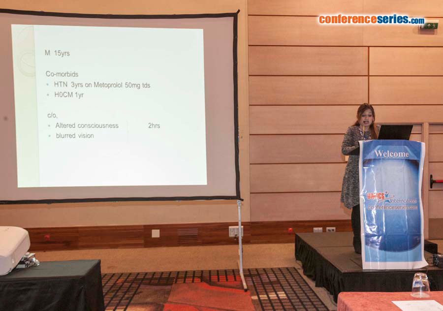
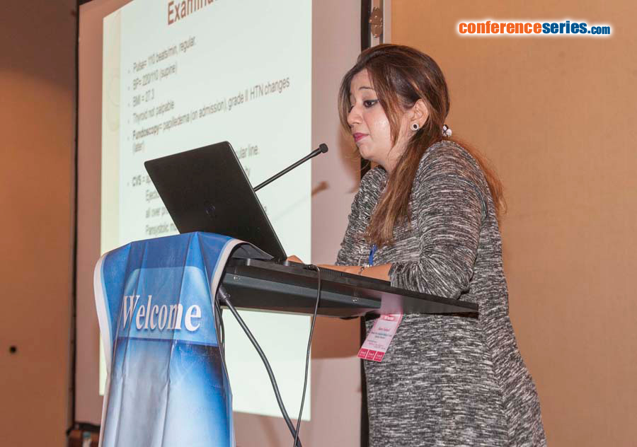
Paraganglioma originates from chromaffin cells of adrenal medulla and autonomic paraganglia, which are derived from the neural crest cells. Paragangliomas are half as common as pheochromocytomas; 69% occurring in head & neck, 22% in abdomen & pelvis and 10% in the thorax. About 70% paragangliomas are sporadic, 30% are hereditary having identifiable germline mutations of Succinate Dehydrogenase Enzyme (SDH). 13 years old male, hypertensive for 3 years, diagnosed as having Hypertrophic Obstructive Cardiomyopathy (HOCM) 1 year back, presented with complaints of altered consciousness and blurring of vision for 2 hours. On examination, blood pressure was 220/110 mmHg. He had a grade III intensity ejection systolic murmur at L lateral sternal border & grade II Pansystolic murmur at mitral area radiating to axilla. GCS was 5/15 with Rt plantar equivocal & Lt plantar up going. Hypertension was persistent in nature with acute elevation occurring especially in morning hours, controlled by intravenous vasodilators during acute elevations and alpha and beta blockers. There was no family history of hypertension. Hormonal workup revealed elevated urinary VMA levels of 68.3 mg/24 hours, suggestive of pheochromocytoma/paraganglioma. Ultrasound and CT-scan of abdomen confirmed large, retroperitoneal soft tissue mass, measuring 6.4×3.5 cm, involving R ureter and R renal vein, invading into the IVC with tumor thrombus with in it. His thyroid function tests, parathyroid hormone (PTH) and calcitonin, ultrasound neck and renal doppler were normal. Echo showed moderate asymmetrically hypertrophied LV and interventricular septum (non-obstructive). A percutaneous biopsy of mass had been done previously and had confirmed paraganglioma. A diagnosis of severe hypertension leading to hypertrophy of myocardium secondary to paraganglioma was made. Tumor was removed in total along with extension in the inferior vena cava from the entrance into R atrium up to R renal vein. Six months post-operative, patient remains well and has resumed his normal day to day activities. He remains normotensive and off all medications. His post-operative urinary VMA levels have also normalized (4 mg/hr). Repeat echocardiographic findings showed regression of changes with only mild asymmetrically hypertrophied left ventricle. This case report together with few others in literature indicates that HOCM like features can be induced by this tumor with complete or partial resolution of these features following successful tumor removal; this in contrast to classic HOCM which is a life-long irreversible disease.